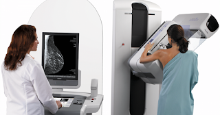
3D Mammography is more effective for breast cancer screening than normal Mammography
Prof.Dr.Dram,profdrram@gmail.com,Gastro Intestinal,Liver Hiv,Hepatitis and sex diseases expert 7838059592,9434143550
 3D mammography, or breast tomosynthesis, is a relatively new breast imaging procedure approved by the U.S. Food and Drug Administration in 2011.Like traditional mammography, 3D mammography uses X-rays to produce images of breast tissue in order to detect lumps, tumors or other abnormalities. 3D mammography is capable of producing more detailed images of breast tissue.Traditional mammography produces just two images of each breast, a side-to-side view and a top-to-bottom view. 3D mammography produces many X-ray images of the breasts from multiple angles to create a digital 3-dimensional rendering of internal breast tissue. This allows radiologists to view the breast in 1-millimeter ‘slices’ rather than just the full thickness from the top and from the side.
3D captures multiple slices of the breast, all at different angles. The images are brought together to create crystal clear 3D reconstruction of the breast. The radiologist is then able to review reconstruction, one slide at a time, almost like turning pages in a book. This makes it easier for doctors to see if there’s anything to be concerned about.
It has been found by a study that in comparison to normal mammorgraphy,3D Mammography better evaluate calcification or altered archtexture of breast in case of suspected sol or fibroadnosis or fibro adenoma and even very useful for detecting lesion in breast with dense tissue.After such eaxmination role of elsatgraphy or MRI has been limited to some extent.It is very useful for screeing women with risk of having breast cancer.
A radiologist will interpret the results of 3D mammography, looking for signs of calcification or masses in the breast tissue, and report findings according to a standardized classification system known as Breast Imaging Reporting and Data System (BI-RADS).
A woman and/or her primary care physician will be notified if her 3D mammography reveals that she has dense breasts or an abnormality in her breast tissue. If an abnormality is detected, radiologist will likely recommend additional imaging procedures to help provide women with greater clarity and determine the cause of any true abnormality or if needed fine needle aspiration cytology or Biopsy for further confirmation.
- Kidney stones universally present hazard in north india,dillution by water prevent it
- Steroid and placebo effect equally for mild persisting asthma with low sputum eosinophils
- Government wants to fix public healthcare staff shortages with ayush docs: will it work?
- Plea in hc for payment of salaries of edmc, north mcd teachers and doctors
- 7 indian pharma companies named in us lawsuit over inflating generic drug prices
- Woman in up dies after explosion in her mouth during treatment,what is diagnosis?
- Woman in up dies after explosion in her mouth during treatment,what is diagnosis?
- Woman in up dies after explosion in her mouth during treatment,what is diagnosis?
- Air pollution ! mothers organising rally in london,anaesthetist choosing gas,will india follow?
- Cardiac arrest is always not sudden as understood -a study

 Comments (
 Category (
 Views (